Recognition of Patterns of Maleficent Objects on Medical Images 2017-10-20T12:44:49+00:00

Recognition of Patterns of Maleficent Objects on Medical Images

Szilvia Nagy

Description

Keywords: medical image; pattern recognition; image processing; colorectal tumor; endoscopy; blood vessels; narrow band imaging

H2020 challenge: Health, demographic change and wellbeing

Knowledge and skills (P: prerequisite; D: desirable, but not necessary): basic knowledge on image taking methods (P); basic knowledge on learning algorithms and soft decision techniques (D); basic image processing (D); basic knowledge on medical imaging (D)

Detecting maleficent medical conditions, especially cancers, is a costly and complicated task. Mostly only a biopsy can determine whether a tumour is cancerous, which causes the patient inconvenience and risk of the operation, as well as extra cost in the medical system. In case of many tumours a non-invasively taken image can also provide information on the nature of the formula, or at least a guidance whether to take biopsy, remove the tumour immediately, or leave it, because the removal is an unnecessary risk in case of a non-maleficent formula.

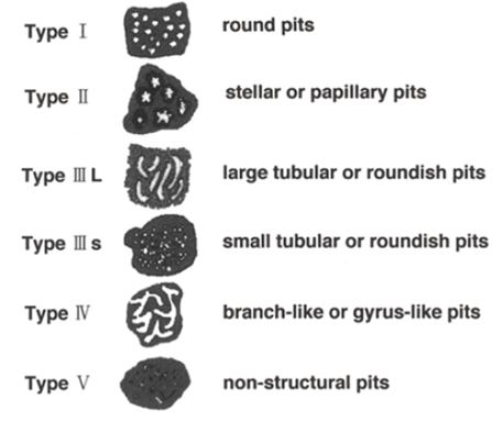
The medical staff is trained to identify maleficent tumours of their specialization field, but an automatic image processing can help their decision, moreover in some cases, like the skin cancers, it could add a tool into the hands of the not specialized persons as well. The non-invasive medical imaging [1][2] contains both the visible light techniques (like endoscopes and microscopes) as well as the higher and lower frequency electromagnetic field based imaging (like X-ray and ultrasound reflection based images). In case of tumours many tell-tale signs can be from the variation of the blood vessel structure around the formula [4-6] to the pattern and colour of the surface of the polyp itself [7-10].

Five INNOSOC students, supervised by two INNOSOC lecturers, will collaborate on answering how ICT can be used in image-based cancer detection. These activities will be conducted as a part of the ERASMUS+ blended mobility and will be finalized during INNOSOC Zagreb 2016 workshop in late April 2016.

Medical image processing is one of positive side effects of the fast proliferation of the ICT into all domains of the society. For example, it can decrease the workload put on medical staff and help them decide in problematic questions or draw attention to smaller details where problems might be present.

Cancer detection is still made by humans, and it will remain so, however, a visual aid can increase effectiveness, and a pre-screening can still be carried out by less qualified personnel and intelligent computer programmes instead of fully trained medical specialists.

The aim of this Case Study is to summarize the technologies used in image-based cancer detection for some types of cancer and compare the applicability of the techniques to the various cases. It is also necessary to determine whether new image processing methods could be used. In most cases the decision about a formula is taken in a crisp, yes or no way, however, many soft decision techniques can also be applied, completed with a learning algorithm, thus it is also necessary to map the applied training algorithms and their efficiency.

Therefore, this Case Study specifically addresses the “Health, demographic change and wellbeing” H2020 challenge.

This Case Study is tightly connected with innovation, intercultural and ICT.

First, the innovation aspect emerges from comparing multiple types of image processing methods used for various purposes that can lead to a common method or a method applied for one type of problem to be applicable in other problems.

Second, although medical image processing is an international problem, acquiring images has different aspects in different cultures. Additionally, interpreting and communicating the results has various cultural aspects as well.

Third, medical images are processed by IT devices, and their transmission has multiple ICT tasks from coding, compressing to videoconferences about the results.

[1] JT Bushberg, JM Boone, The essential physics of medical imaging, (Wolters Kluwer, Philadelphia 2012).

[2] J Beutel, HL Kundel, RL Van Metter, Handbook of Medical Imaging, (2000, Society of Photo-Optical Instrumentation Engineers)

[3] J. J. W. Tischendorf, H. E. Wasmuth, A. Koch, H. Hecker, C. Trautwein, and R. Winograd, “Value of magnifying chromoendoscopy and narrow band imaging (NBI) in classifying colorectal polyps: a prospective controlled study”, Endoscopy, Volume 39, Thieme, Stuttgart-New York, 2007, pp. 1092-1096.

[4] Robert Folberg, Volker Rummelt, Rita Parys-Van Ginderdeuren, Taekyu Hwang, Robert F. Woolson, Jacob Pe’er, Lynn M. Gruman, The Prognostic Value of Tumor Blood Vessel Morphology in Primary Uveal Melanoma, Ophthalmology, Vol. 100, pp 1389–1398 (1993)

[5] Rakesh K. Jain, Determinants of Tumor Blood Flow: A Review, Cancer Res, Vol 48; p. 2641, (1988)

[6] K. Søreide, B.S. Nedrebø, A. Reite et al., „Endoscopy Morphology, Morphometry and Molecular Markers: Predicting Cancer Risk in Colorectal Adenoma”, Expert Rev. Mol. Diagn, vol. 9, pp. 125-137, 2009.

[7] S. Kudo, S. Hirota, T. Nakajima, et al., “Colorectal tumours and pit pattern”. J Clin Pathol, vol. 47, pp.880-885, 1994.

[8] S. Kudo, S. Tamura, T. Nakajima, et al. Diagnosis of colorectal tumorous lesions by magnifying endoscopy. Gastrointest Endosc, vol. 44, pp. 8-14, 1996.

[9] S. Kudo, C.A. Rubio, C.R., Teixeira, et al. Pit pattern in colorectal neoplasia: endoscopic magnifying view. Endoscopy, vol. 33, pp. 367-373, 2001.

[10] J. R. Jass, “Classification of colorectal cancer based on correlation of clinical, morphological and molecular features”, Histopathology, Volume 50, Wiley, 2006, pp. 113–130.

[11] I. Rácz, M. Jánoki, and H. Saleh, “Colon Cancer Detection by ‘Rendezvous Colonoscopy’: Successful Removal of Stuck Colon Capsule by Conventional Colonoscopy”, Case Rep. Gastroenterol., Volume 4, Karger, 2010, pp. 19–24.

[12] Rozenn Dahyot, Fernando Vilarino, and Gerard Lacey, ,,Improving the Quality of Color Colonoscopy Videos”, Hindawi Publishing Corporation, EURASIP Journal on Image and Video Processing.

[13] Vipul Sharan, Naveen Keshari, Tanay Mondal, Biomedical Image Denoising and Compression in Wavelet using MATLAB, International Journal of Innovative Science and Modern Engineering (IJISME)
Blood vessels around a kidney cancer (from the online cover of Science Translational Medicine, Vol 3, Issue 66 – credit: C. Bickel / Science Translational Medicine)
Colorectal polyp pit patterns (from: Nikolas Eleftheriadis, Haruhiro Inoue, Haruo Ikeda, Manabu Onimaru, Akira Yoshida, Roberta Maselli, Grace Santi, Shin-ei Kudo, “Definition and Staging of Early Esophageal, Gastric and Colorectal Cancer”, Journal of Cancer, Vol. 2, pp. 161-178 (2014))

Questions that need answers

  • What are image acquiring methods used in tumour classification?
  • Are there specially developed imaging techniques for some aspects of tumour detection (e.g., for recognition of surfaces and interfaces of tumours or for enhancing the visibility of the blood vessels)?
  • What are the most lethal cancer types in Europe and in the other continents?
  • What is the usual diagnose method for the most lethal tumours?
  • What type of image processing methods are used in detection or classification of the most dangerous tumour types?
  • What type of soft or crisp decision techniques are used in tumour classification? What types of evolutionary or learning algorithms are applied?
  • How the IT classification success rate is related to the human classification success rate or the biopsy results? What tumour types are well classifiable and what are hard to determine? Why (from image processing point of view)?

Case study supervisors

Szilvia Nagy does not have a photo :(

Szilvia Nagy

Lecturer

Case study students

Sandra Yuste Muñoz does not have a photo :(

Sandra Yuste Muñoz

INNOSOC 2016 & 2017 Student

Georgi Nikolov does not have a photo :(

Georgi Nikolov

INNOSOC 2016 Student

Irena Oršolić does not have a photo :(

Irena Oršolić

INNOSOC 2016 Student

József Zákány does not have a photo :(

József Zákány

INNOSOC 2016 Student

Multilingual Case Studies

United Kingdom    

Bulgaria    

Germany    

Spain    

France    

Croatia    

Hungary    

Romania    

Slovakia    

Skip to content